Phase 1: Understanding the Carpal Tunnel and Median Nerve Compression
The carpal tunnel is a narrow, rigid passageway at the base of your palm. The "floor" and "walls" of this tunnel are formed by small wrist bones (carpal bones), while the "roof" is formed by a tough, unyielding band of connective tissue called the transverse carpal ligament.
Through this tight space pass exactly 10 structures: nine flexor tendons (which bend your fingers) and one crucially important nerve—the Median Nerve. The median nerve provides feeling to your thumb, index, middle, and half of the ring finger, while also controlling the muscles at the base of your thumb.
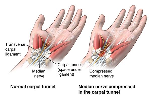
Carpal Tunnel Syndrome (CTS) occurs when the flexor tendons swell (tenosynovitis), fluid accumulates in the tunnel, or the tissues degenerate, increasing the pressure inside this closed space. Because the roof (the ligament) cannot expand, the pressure ultimately squeezes the soft, vulnerable median nerve. This compression restricts blood flow to the nerve (ischemia), leading to the classic symptoms of numbness, tingling, and pain.
Phase 2: Diagnostic Evaluation & Clinical Red Flags
Patients classically report waking up in the middle of the night with severe numbness or a "pins-and-needles" sensation in their hand. A hallmark symptom is the "Flick Sign"—patients instinctively shake or flick their hands to try and restore circulation and sensation. As the condition worsens, clumsiness, dropping objects, and difficulty performing fine motor tasks (like buttoning a shirt) become common.
Clinical Examination: Diagnosis is heavily reliant on physical provocation tests in the clinic:
- Durkan's Test (Carpal Compression Test): Direct manual pressure is applied over the carpal tunnel for 30 seconds. This is considered the most sensitive and specific physical test for CTS, rapidly reproducing the numbness.
- Phalen's Maneuver: The patient holds their wrists in complete, unforced flexion for 60 seconds, which temporarily increases pressure in the tunnel.
- Tinel's Sign: Light tapping over the median nerve at the wrist elicits a "shock-like" tingling sensation shooting into the fingers.
- Visual Inspection: In severe, chronic cases, we look for Thenar Atrophy—a visible wasting (shrinkage) of the muscle pad at the base of the thumb. This is a red flag indicating severe nerve damage.
Advanced Diagnostics: To definitively confirm the diagnosis and determine the severity of nerve damage, a Nerve Conduction Study (NCS) and Electromyography (EMG) are considered the gold standard. These tests measure how fast and how well electrical signals travel down the median nerve.
Phase 3: First-Line Conservative Protocols (Splinting)
If caught early, mild to moderate Carpal Tunnel Syndrome can be effectively managed without surgery by addressing the mechanical compression of the nerve.
Neutral Wrist Splinting: The foundation of conservative care is wearing a rigid wrist brace, particularly at night. It is critical that the splint holds the wrist in a neutral (straight) position, not bent backward (extended). Medical studies show that pressure inside the carpal tunnel is lowest when the wrist is perfectly straight. Because people naturally bend their wrists while sleeping, nighttime splinting prevents the hours-long nerve compression that causes morning pain.
Ergonomics and Activity Modification: Repetitive gripping, forceful wrist flexion, and prolonged use of vibrating power tools exacerbate CTS. Adjusting keyboard and mouse setups so the wrist hovers in neutral, taking frequent breaks, and performing nerve-gliding exercises (to help the median nerve slide freely within the tunnel) are highly beneficial.
Phase 4: Medical Management & Corticosteroid Injections
When splinting and ergonomic modifications fail to provide adequate relief, we utilize targeted medical interventions to aggressively lower the pressure within the tunnel.
Corticosteroid Injections: A highly localized injection of a corticosteroid into the carpal tunnel is an excellent treatment for moderate CTS. It dramatically reduces the inflammatory swelling (tenosynovitis) of the tendon sheaths surrounding the nerve, thereby freeing up space.
Injections are often performed under ultrasound guidance to ensure the medication is placed perfectly around the nerve without injuring it. While injections provide rapid and profound relief—and can be diagnostic in unclear cases—studies show that in roughly 50% to 70% of patients, symptoms may recur within a year, eventually necessitating surgery.
Note: Oral medications like NSAIDs (ibuprofen) or Vitamin B6 have shown very limited, if any, long-term efficacy in treating the mechanical compression of CTS.
Phase 5: Surgical Indications & Carpal Tunnel Release
Surgery is strongly indicated for patients who fail conservative treatment after 3 to 6 months, those who have intolerable night pain, and strictly required for patients showing "red flags": constant (unrelenting) numbness, muscle weakness, or visible thenar muscle atrophy on EMG.
Carpal Tunnel Release (CTR): The surgical goal is brilliantly simple: completely divide (cut) the transverse carpal ligament to open the roof of the tunnel. This permanently increases the volume of the carpal tunnel by up to 24%, instantly relieving the pressure on the median nerve.
- Open Release: A small, 1.5 to 2-inch incision is made in the palm. The surgeon directly visualizes the ligament and cuts it, ensuring the nerve is safely protected.
- Endoscopic Release: A camera (endoscope) is inserted through a tiny incision at the wrist crease, and the ligament is cut from the inside. This often results in less early palm pain, though both techniques yield identical, excellent long-term outcomes.
Phase 6: Post-Operative Recovery & Nerve Healing
Carpal Tunnel Release is one of the most successful and reliable procedures in orthopaedic surgery, with patient satisfaction rates exceeding 90%.
Immediate Relief vs. Prolonged Healing: Most patients report that their frustrating nighttime waking and nerve pain vanish almost immediately after surgery. However, the timeline for the return of normal sensation (numbness resolving) and grip strength depends entirely on how badly the nerve was crushed before surgery. A heavily damaged nerve may take 6 to 12 months to fully regenerate.
"Pillar Pain": It is common to experience deep aching at the base of the palm (on the sides of the incision) for 1 to 3 months post-operatively. This is known as "pillar pain" and resolves as the divided ligament heals in a lengthened, relaxed position. Gentle scar massage and early finger mobilization are encouraged to prevent internal scarring.
Authentic Sources & Clinical References
The medical protocols and data discussed in this patient guide are grounded in high-level orthopaedic research and peer-reviewed literature. Key reference sources include:
1. American Academy of Orthopaedic Surgeons (AAOS). "Management of Carpal Tunnel Syndrome Evidence-Based Clinical Practice Guideline." Published 2016.
2. Gelberman RH, Hergenroeder PT, Hargens AR, Lundborg GN, Akeson WH. "The carpal tunnel syndrome. A study of carpal canal pressures." The Journal of Bone & Joint Surgery (American). 1981;63(3):380-383.
3. Marshall S, Tardif G, Ashworth N. "Local corticosteroid injection for carpal tunnel syndrome." Cochrane Database of Systematic Reviews. 2007;(2):CD001554.
4. Gerritsen AA, de Krom MC, Struijs MA, Peters RJ, Harderwijk AL, Bouter LM. "Conservative treatment of carpal tunnel syndrome: a systematic review of randomized controlled trials." Journal of Neurology. 2002;249(3):272-280.