Understanding the Torn ACL: Why Stability Matters
The Anterior Cruciate Ligament (ACL) is the primary stabilizer of the knee joint. It prevents the tibia (shin bone) from sliding forward abnormally relative to the femur (thigh bone) and resists excessive rotational torque when pivoting, decelerating, or landing from a jump.
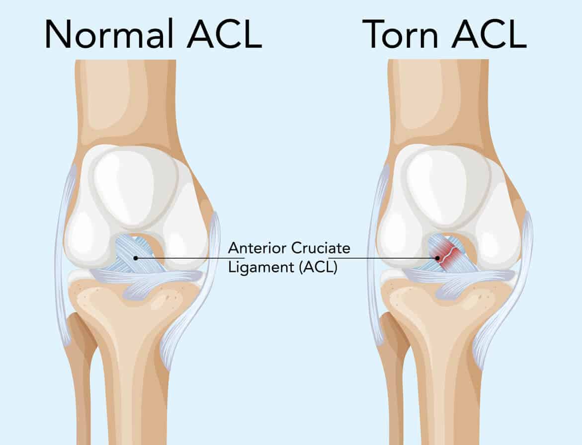
Unlike extra-articular collateral ligaments (such as the MCL), a completely torn ACL cannot heal on its own. The joint is immersed in synovial fluid, which washes away the biological blood clot needed to bridge a torn ligament. Leaving a ruptured ACL untreated in active individuals causes recurrent episodes of knee "giving way" (subluxation), leading to irreversible damage to the shock-absorbing meniscal cushions and early-onset osteoarthritis.
Hallmark Symptoms & Clinical Signs:
- An Audible "Pop" at Injury: Most patients report hearing or feeling a distinct snap inside the knee during a sudden non-contact pivot, sidestep, or awkward jump landing.
- Rapid Joint Hemarthrosis: Sudden, tense knee swelling that develops within 2 to 4 hours post-injury due to bleeding from the ruptured middle genicular artery.
- Inability to Bear Weight Immediately: Acute instability accompanied by localized pain and temporary loss of terminal knee extension.
- Pivot-Shift & Positive Lachman: Clinical examination demonstrates abnormal anterior tibial translation (positive Lachman test) and rotational laxity (positive pivot-shift test), confirmed accurately via high-resolution 1.5T/3T Knee MRI.
Anatomic Arthroscopic Reconstruction: The Keyhole Approach
ACL surgery is performed using minimally invasive keyhole arthroscopy. Through two or three 5 mm portal incisions, a high-definition 4K arthroscope and specialized micro-instruments are introduced into the knee joint. The damaged, frayed remnants of the native torn ligament are gently debrided while preserving mechanoreceptor-rich tissue stumps.
True Anatomic Footprint Placement: Modern orthopaedic sports surgery rejects old-fashioned "non-anatomical" trans-tibial drilling. We utilize an independent transportal or retro-reaming technique. This allows millimeter-level drilling of the femoral tunnel precisely within the native ACL footprint on the lateral femoral condyle. Reproducing the native oblique footprint restores both anteroposterior stability and rotational pivoting resistance, minimizing long-term graft failure rates.
Secure Biological Tunnel Ingrowth: The reconstructed graft is routed through precision femoral and tibial bone tunnels. Over an 8 to 12-week biological process known as ligamentization, new blood vessels and Sharpey-like collagen fibers anchor the tendon graft into host bone, converting it into a living, responsive intra-articular ligament.
Selecting the Right Autograft: Tailored to Your Demands
Because synthetic ligaments have historically shown high failure rates, autologous tissue (the patient's own tissue) remains the international gold standard. We evaluate age, athletic goals, occupational demands, and joint laxity to choose the optimal graft:
| Autograft Type | Biomechanical Advantages | Primary Clinical Indication |
|---|---|---|
| Quadrupled Hamstrings (ST/G) | Minimal anterior incision; low donor-site morbidity; robust tensile strength exceeding native ACL. | Recreational athletes, running enthusiasts, desk professionals, and active daily lifestyles. |
| Peroneus Longus (PLT) | Large graft diameter (often >8.5 mm); completely spares knee flexors and hamstring mechanics. | Fast bowlers, sprinters, martial artists, and individuals with naturally small hamstring tendons. |
| Bone-Patellar Tendon-Bone (BTB) | Bone-to-bone healing in bone tunnels; earliest biological incorporation; proven gold standard. | High-level contact sports athletes (football, rugby) requiring rapid structural bone-tunnel union. |
| Quadriceps Tendon Autograft | Predictably thick diameter, excellent stiffness profile, and minimal donor-site kneeling tenderness. | Revision ACL surgery or multi-ligament reconstructions requiring substantial collagen volume. |
Time-Tested Imported Implants: Why We Never Compromise
In ACL reconstruction, your new ligament graft is completely dependent on mechanical fixation devices until bone ingrowth occurs over the first 8 to 12 weeks. If a fixation button slips by even 1 to 2 millimeters or an interference screw cuts through the tendon fibers, the graft loses tension, causing residual laxity or surgical failure.
For this reason, we exclusively use time-tested, internationally proven imported implants manufactured by global leaders in sports medicine. We strictly refuse unverified local copies or untested hardware.
Global Pioneer Brands We Routinely Deploy
These systems feature decades of peer-reviewed biomechanical validation, minimal cyclic displacement under load, and unmatched tensile yield strength:
| Device Type | Time-Tested Imported Systems | Clinical Superiority & Purpose |
|---|---|---|
| Femoral Suspensory Fixation | Arthrex TightRope® RT/ABS Smith & Nephew ULTRABUTTON® |
Adjustable-loop cortical buttons providing four-point locking mechanisms. Eliminates loop elongation and protects the graft from abrasive sawing forces during tensioning. |
| Tibial Aperture Fixation | Arthrex BioComposite™ / FastThread™ Smith & Nephew BIOSURE™ REGENESORB™ |
Osteoconductive biocomposite interference screws (PLDLA + β-TCP / Calcium Phosphate). Gradually absorbs over time, promoting genuine bone ingrowth into the tunnel rather than leaving permanent voids. |
| Meniscal Repair Devices | Smith & Nephew FAST-FIX™ 360 Arthrex FiberStitch™ |
All-inside low-profile meniscal suturing implants with pre-tied 2-0 ultra-high-molecular-weight polyethylene (UHMWPE) FiberWire® sutures for anatomical meniscal salvage. |
Using premium imported implants ensures the graft is held under calibrated physiological tension from Day 1. This allows early weight-bearing, rapid quadriceps activation, and safe accelerated rehabilitation without fear of implant failure.
Meniscus Salvage & Lateral Extra-Articular Tenodesis (LET)
Over 50% of acute ACL tears involve concomitant damage to the medial or lateral meniscus. In the past, torn meniscus portions were routinely trimmed away (meniscectomy). Today, our philosophy is to save the meniscus whenever biologically possible:
- All-Inside & Inside-Out Meniscal Repair: Utilizing specialized low-profile suture devices to stitch vascular "red-red" and "red-white" zone meniscal tears. Preserving the meniscus preserves native shock absorption and halts premature joint wear.
- Addressing Meniscal Ramp Lesions: Hidden tears of the posterior horn of the medial meniscus attachment are routinely inspected via posteromedial trans-notch viewing and sutured to restore rotational control.
- Lateral Extra-Articular Tenodesis (Modified Lemaire / ALL Reconstruction): In patients with high-grade rotational pivot shift, generalized ligamentous laxity (hyperextension), or young athletes returning to pivoting sports, we augment the ACL with a simple lateral band tenodesis. Clinical studies show this reduces graft re-rupture rates by more than 50% by shielding the internal ACL graft from high rotational torque.
Operating Room Sterility: Protecting Your Reconstructed Graft
In sports arthroscopy, maintaining total intra-operative sterility is critical. While arthroscopic incisions are small, an autograft placed into bone tunnels functions as an avascular tissue scaffold until it revascularizes over several months. Preventing bacterial colonization is vital for graft survival and joint safety.
We perform all ACL reconstructions exclusively in accredited hospitals with modern infection-control systems and dedicated orthopaedic teams:
Multi-Layered Surgical Protection Protocols
- Laminar Airflow Operating Theatres: Continuous positive-pressure HEPA air filtration keeping the operative zone free of airborne micro-particulates.
- Central Sterile Services (CSSD): Multi-stage monitored autoclave and plasma sterilization of precision arthroscopic instrumentation and drill guides.
- Graft Preparation Protocol: Dedicated graft-preparation stations with sterile vancomycin presoaking, proven in orthopaedic trials to reduce joint infection risks.
- Dedicated Arthroscopy Nursing Teams: Trained scrub nurses and technicians adhering strictly to sterile fluid management and drape integrity.
Maintaining these hospital infrastructure standards and using time-tested imported implants naturally impacts overall surgical package pricing compared to non-sterile basic facilities. However, this investment safeguards your knee from devastating joint sepsis, prevents graft lysis, and ensures the safest path back to sport.
Phased Sports Rehabilitation: The Journey to Return to Play
Surgery accounts for only half the battle; the remaining 50% depends on biology-driven rehabilitation. We employ objective criteria rather than arbitrary calendar dates to guide progression:
| Rehab Phase | Primary Milestones & Goals | Permitted Activities |
|---|---|---|
| Phase 1 (Weeks 0–4) | Full knee extension (0°), effusion control, quad reactivation, patellar mobility. | Walker/crutch weaning, static quad sets, straight-leg raises, stationary cycle rocking. |
| Phase 2 (Weeks 5–12) | Full knee flexion, normal gait, single-leg closed kinetic chain strength. | Full stationary cycling with resistance, leg press, mini-squats, step-ups, swimming (flutter kick). |
| Phase 3 (Months 3–6) | Restoring symmetrical quadriceps/hamstring muscle bulk and dynamic balance. | Outdoor straight-line jogging (on flat track), agility ladder drills, low-level plyometrics. |
| Phase 4 (Months 6–9+) | Sport-specific deceleration, multi-directional cutting, rotational control. | Cutting drills, sport-specific skill practice, non-contact team training sessions. |
| Return to Play (Months 9–12) | Limb Symmetry Index (LSI) >90% on single-leg hop testing, psychological readiness (ACL-RSI). | Full competitive clearance for cricket, football, hockey, martial arts, and high-intensity athletics. |
Surgery Costs & Cashless Insurance in Jamshedpur
The overall package cost of arthroscopic ACL surgery depends on chosen imported graft fixators (suspensory buttons vs. bioabsorbable screws), whether concurrent meniscal repair is performed, and selected hospital room categories.
Comprehensive Cashless Insurance Support
We cover almost all major private health insurance policies, corporate TPAs, and recognized health panels.
Because policy terms, sport injury exclusion clauses, room-rent capping, and waiting periods differ widely across insurers, please share your policy card directly via WhatsApp prior to admission to check your eligibility and ensure smooth cashless pre-authorization.
Confirm Insurance on WhatsAppOur clinical team provides transparent pre-operative counseling covering implants, OT disposable kits, arthroscopic shaver blades, radiofrequency probes, and initial post-op bracing.
Frequently Asked Questions About ACL Tears
Why do you only use imported ACL implants?
Implant failure in ACL surgery typically manifests as suture breakage or loop elongation during everyday walking. Time-tested imported systems from Arthrex, Smith & Nephew, and Stryker have undergone extensive cyclic testing and registry validation, ensuring zero slippage and reliable biological union.
Can I delay ACL surgery, and will it damage my knee?
A short delay of 2 to 3 weeks until acute post-injury swelling settles and range of motion returns ("pre-habilitation") is recommended. However, indefinitely delaying surgery while returning to pivoting sports carries an elevated risk of meniscal tears and joint cartilage damage.
How long will I need crutches after surgery?
For an isolated ACL reconstruction, patients typically use elbow crutches for 10 to 14 days for gait protection. If a meniscal repair or cartilage grafting is performed concurrently, crutch use and partial weight-bearing are maintained for 4 to 6 weeks to protect the repaired tissue.
Does harvesting the hamstring or peroneus tendon cause permanent weakness?
No. When harvested carefully using specialized tendon strippers, the body adapts through hypertrophic compensation of the adjacent synergistic muscles. Long-term functional studies show no noticeable deficit in daily walking or sports performance.
What are the chances of an ACL re-tear after surgery?
Modern anatomical ACL reconstructions boast a 90% to 95% success rate. Re-tear risks are highest in young athletes returning to sport too early (under 9 months) before full graft "ligamentization" and neuromuscular control have matured. Concomitant LET and disciplined physical therapy significantly reduce this risk.
Clinical References & Literature
1. van Melick N, et al. "Evidence-based clinical practice update: practice guidelines for anterior cruciate ligament rehabilitation based on a systematic review and multidisciplinary consensus." British Journal of Sports Medicine. 2016;50(24):1506-1515.
2. Getgood AMJ, et al. "Lateral Extra-articular Tenodesis Reduces Failure of Hamstring Tendon Autograft Anterior Cruciate Ligament Reconstruction: 2-Year Outcomes From the STABILITY Study Randomised Clinical Trial." The American Journal of Sports Medicine. 2020;48(2):285-297.
3. Perez-Prieto D, et al. "Presoaking ACL grafts in vancomycin reduces post-operative infection: a systematic review and meta-analysis." Knee Surgery, Sports Traumatology, Arthroscopy. 2018;26(10):3139-3146.