Understanding Calcaneus Fractures: The Heel Bone Shock Absorber
The calcaneus (heel bone) is the largest tarsal bone in the foot, serving as the primary weight-bearing foundation and lever arm for walking. Calcaneus fractures typically result from high-energy axial loading—such as falling from a height (ladder, roof, scaffolding) or severe motor vehicle accidents. The talus bone acts like a wedge, smashing down into the calcaneus and flattening its structure, destroying the subtalar joint and reducing Bohler's angle (the crucial radiographic measurement of heel height).
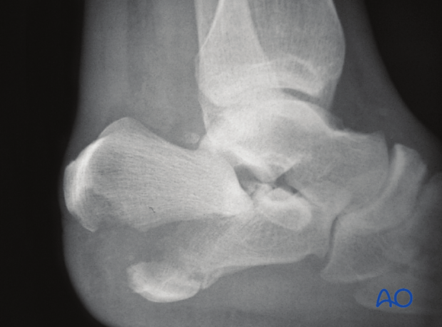
Without proper anatomical reconstruction, patients suffer from permanent heel widening, loss of height, chronic pain, sub-talar arthritis, and difficulty fitting into standard footwear.
Tongue-Type Calcaneus Fractures: A Soft-Tissue Emergency
A tongue-type calcaneal fracture is a distinct injury pattern where a large posterior bone fragment containing the Achilles tendon attachment breaks off as a single unit and tilts backward.
Why This Is an Emergency: The sharp anterior tip of this displaced tongue-like fragment acts like a blade pointing directly toward the overlying heel skin from the inside. If left unreduced, it can rapidly cause skin tenting, pressure ischemia, and full-thickness skin necrosis (sloughing), turning a closed fracture into an open, infected wound. Urgent reduction and percutaneous or open pin/screw fixation is required to drop the fragment back down and relieve skin tension.
Conservative Casting vs. Surgery: Treatment Decisions
Determining whether a broken heel requires an operation depends on joint surface step-offs, Bohler's angle collapse, and skin condition.
When Conservative Cast Care Works:
- Non-Displaced or Minimally Displaced Fractures: Intra-articular step-offs are under 2 mm, and Bohler's angle is fully preserved (20 to 40 degrees).
- Medically High-Risk Patients: Severe vascular disease, active smokers with extreme skin necrosis risks, or frail patients with low functional demands.
When Surgical Fixation (ORIF or Percutaneous Pinning) Is Mandatory:
- Displaced Intra-Articular Fractures: Articular displacement > 2 mm across the posterior facet of the subtalar joint.
- Collapsed Bohler's Angle: Flattened or negative heel angles resulting in loss of propulsive power and flatfoot deformity.
- Tongue-Type Fractures with Skin Tenting: Urgent fixation required to prevent skin breakdown.
Surgical Techniques: Extended Lateral ORIF vs. Sinus Tarsi Approach
Depending on swelling, skin condition, and fracture geometry, modern trauma surgery utilizes advanced approaches:
| Surgical Modality | Technique & Approach | Best Indicated For |
|---|---|---|
| Extended Lateral ORIF | Full-thickness L-shaped lateral incision providing direct visualization of the entire subtalar joint and lateral wall. | Shattered, highly comminuted intra-articular calcaneal fractures requiring plate scaffolding. |
| Minimally Invasive Sinus Tarsi Approach | A small 3–4 cm incision over the outer ankle, protecting soft tissues and lowering wound complication rates. | Displaced intra-articular fractures with moderate comminution. |
| Percutaneous Screw Fixation / Reduction | Closed manipulation with leverage pins (Essex-Lopresti technique) secured with cannulated screws. | Acute tongue-type calcaneal fractures requiring urgent soft-tissue rescue. |
Risks of Inadequate Treatment: Subtalar Arthritis & Flatfoot
Failing to restore heel height and subtalar joint congruence leads to permanent disability:
- Post-Traumatic Subtalar Arthritis: Unresolved cartilage steps cause grinding beneath the ankle bone, resulting in severe pain when walking on uneven ground.
- Heel Widening & Peroneal Impingement: A widened calcaneus rubs against the outer fibula bone and peroneal tendons, causing chronic lateral ankle pain and clicking.
- Traumatic Flatfoot (Valgus Deformity): Loss of Bohler's angle flattens the arch, altering total lower limb biomechanics.
Time-Tested Imported Implants: Calcaneal Locking Plates
The calcaneus is subjected to severe multidirectional compressive forces during heel strike. Standard plates lack the multi-planar screw trajectories required to capture fragmented bone.
Global Pioneer Systems Deployed at Our Center
We deploy certified, time-tested imported titanium calcaneal locking plate systems and cannulated screw sets:
These plates feature anatomical contouring and divergent locking screw holes that cradle the subtalar joint surface and secure the posterior tuberosity effectively.
Operating Room Sterility & Delicate Soft-Tissue Management
Heel skin has poor microvascular circulation compared to other parts of the body. Preventing wound edge necrosis and deep infection is the most critical surgical challenge.
Multi-Layered Soft-Tissue & Infection Control Protocols
- Swelling Timing Protocols: Elective ORIF is typically scheduled once skin wrinkling returns (usually 5 to 10 days post-injury) to minimize wound breakdown.
- Laminar Airflow Operating Theatres: Ultra-clean positive-pressure HEPA air filtration safeguarding deep foot reconstructions against contamination.
- Atraumatic Retraction: Gentle handling of full-thickness skin flaps during plate insertion to preserve local blood supply.
Phased Recovery Timeline: From Non-Weight Bearing to Normal Walking
Calibrated fixation provides stable internal alignment, allowing structured rehabilitation while biological bone union progresses:
| Timeframe | Bone Healing & Soft-Tissue Status | Permitted Foot & Ankle Function | Physiotherapy & Rehabilitation Focus |
|---|---|---|---|
| Week 1 to 6 | Incision heals cleanly; calcaneal plate secures heel structure; strict non-weight bearing. | Resting foot elevated; using a wheelchair, knee scooter, or crutches with zero weight on heel. | Active ankle pumps, toe wiggles, and knee/hip range of motion exercises. |
| Weeks 6 to 12 | Early bridging bone callus visible across calcaneal fragments on checkup X-rays. | Transitioning into a removable walking boot; starting touch-down to partial weight bearing (15–25 kg). | Gentle subtalar inversion-eversion stretching and pool therapy. |
| Weeks 12 to 16 | Solid radiographic bone union and cortical consolidation confirmed. | Transitioning from boot to supportive supportive athletic shoes; progressing to full weight bearing. | Proprioceptive balance training, calf raises, and gait retraining. |
| Months 4 to 6 | Complete structural remodeling of the heel architecture. | Unrestricted walking, stair climbing, driving, and return to moderate work duties. | Full functional stamina, endurance building, and return to active lifestyle. |
Surgery Costs & Cashless Mediclaim in Jamshedpur
Calcaneus fracture surgery packages depend on the fracture classification (tongue-type emergency vs. comminuted intra-articular break), the selection of imported titanium locking plates, and the hospital room category selected.
Emergency Cashless Insurance & TPA Support
We cover almost all major private health insurance policies, corporate TPAs, and recognized health panels.
Because acute accidental heel fractures resulting from falls from height are treated as medical emergencies, most health policies cover hospitalization without standard waiting periods. Families should share the patient's insurance card and accident details directly on our clinic WhatsApp at +91 7739798850 for immediate pre-authorization desk assistance.
Confirm on whatsappOur administrative desk ensures transparent, upfront billing covering imported titanium plate costs, OT consumables, anesthesia fees, and follow-up care with zero hidden surprises.
Frequently Asked Questions by Patients & Families
What is a tongue-type calcaneus fracture?
A tongue-type calcaneal fracture is a specific displacement pattern where a single large bone fragment containing the posterior articular surface and Achilles tendon insertion is sheared off and tilted backward. Because this sharp fragment can push outward and threaten the heel skin from the inside, it is treated as an orthopaedic emergency requiring prompt reduction to prevent skin necrosis.
Why is Bohler's angle important in heel bone fractures?
Bohler's angle is the key radiographic measurement used to assess the structural height and biomechanical integrity of the heel bone. Restoring this angle during surgery ensures normal walking mechanics, proper calf muscle tension, and prevents permanent flatfoot deformity.
Can a broken heel bone heal without surgery?
Non-operative casting is only considered for non-displaced fractures where the subtalar joint surfaces and Bohler's angle are completely preserved. Displaced intra-articular fractures and emergency tongue-type breaks require surgical fixation to prevent chronic pain, subtalar arthritis, and shoe-fitting difficulties.
How long does it take to walk after calcaneus surgery?
Bone healing typically requires 10 to 12 weeks. Patients remain non-weight bearing in a splint or boot for the initial 6 to 8 weeks to allow secure bone healing and soft-tissue recovery, gradually progressing to partial and full weight bearing over 3 to 4 months.
Is calcaneus fracture surgery covered under cashless health insurance in Jamshedpur?
Yes. Acute traumatic heel fractures resulting from falls from height or road accidents qualify as emergency surgical admissions covered under private health insurance policies and corporate TPAs without waiting periods.
Clinical References & Scientific Evidence
1. Essex-Lopresti P. "The mechanism, reduction technique, and results in fractures of the os calcis." British Journal of Surgery. 1952;39(157):395-419.
2. Rüedi TP, Buckley RE, Moran CG. "AO Principles of Fracture Management." AO Foundation / Thieme. 2007.
3. Buckley R, et al. "Operative compared with nonoperative treatment of displaced intra-articular calcaneal fractures: a prospective, randomized, controlled multicenter trial." The Journal of Bone & Joint Surgery (Am). 2002;84(10):1733-1744.