Meniscus Tears & Mechanical Symptoms: Understanding Joint Mechanics
The medial and lateral menisci are crescent-shaped, specialized fibrocartilaginous pads positioned between the femoral condyle (thigh bone) and the tibial plateau (shin bone). Rather than simple inert spacers, they are sophisticated shock absorbers that transmit up to 70% of total axial joint loads, lubricate articular cartilage, and provide secondary anterior-posterior joint stability.
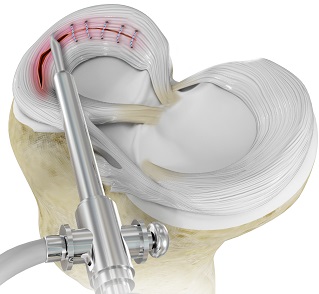
Meniscus tears occur along two distinct clinical paths in Jamshedpur: acute traumatic tears in athletes and industrial workers (often associated with an ACL rupture or high-velocity twisting) and degenerative cleavage tears in individuals over age 40 from repetitive deep squatting or cartilage dehydration.
Hallmark Symptoms of a Clinically Significant Meniscus Tear:
- Mechanical Joint Locking: The knee physically jams or locks at a specific angle (often 30° to 45° of flexion) due to an unstable "bucket-handle" fragment flipping into the intercondylar notch.
- Audible Clicking & Catching: A painful pop or snapping sensation felt along the joint line when rising from a chair or pivoting on a planted foot.
- Localized Joint Line Tenderness: Pinpoint, sharp pain directly along the medial (inner) or lateral (outer) joint line margin on clinical palpation.
- Delayed Effusion / Swelling: Unlike the rapid hemarthrosis of an ACL tear, isolated meniscal tears typically produce recurrent, dull swelling 12 to 24 hours after weight-bearing activity.
The "Save the Meniscus" Philosophy: Halting Early Osteoarthritis
Historically, torn meniscus tissue was regarded as expendable, and surgeons routinely performed a complete meniscectomy (cutting away the entire cushion). Decades of international registry follow-up have revealed the devastating consequence: removing the meniscus results in a 200% to 300% surge in peak contact stresses on the cartilage, frequently causing severe Grade IV osteoarthritis in young patients within 10 to 15 years.
Our clinical approach is anchored in meniscal preservation and biological salvage whenever possible:
- Red-Red Zone (Outer 1/3rd): High vascularity supplied by the perimeniscal capillary plexus; heals with high predictability when anatomical apposition is restored.
- Red-White Zone (Middle 1/3rd): Intermediate blood supply; responds favorably to all-inside or outside-in suturing combined with biological trephination (micro-needling to induce healing clots).
- White-White Zone (Inner 1/3rd): Truly avascular; reserved for ultra-conservative, selective micro-contouring (partial meniscectomy) only when structural repair is biologically unviable.
Advanced Arthroscopic Repair Techniques: All-Inside & Inside-Out
Meniscus repair is performed 100% keyhole through two tiny 5 mm portals under 4K optical visualization. Specialized techniques are matched to tear configuration:
All-Inside Meniscal Suture Systems: Ideal for posterior horn and mid-body tears. Utilizing low-profile articulating needles, tiny polyetheretherketone (PEEK) or all-suture anchors are deployed on the outer capsule. Pre-tied sliding knots (such as 2-0 UHMWPE sutures) compress the tear margins with controlled tension without requiring extra incisions.
Inside-Out Suturing for Large Bucket-Handle Tears: Considered the gold standard for large, complex bucket-handle tears spanning the posterior and middle zones. Long double-loaded flexible needles pass from inside the joint out through a protected mini-incision, placing vertical mattress sutures every 3 to 4 millimeters for maximum mechanical grip.
Biological Enhancement: To maximize healing rates in avascular zones, we routinely perform joint abrasion, parameniscal trephination (channelling), and bone marrow venting to release rich growth factors and autologous mesenchymal stem cells into the repair bed.
The Outside-In Repair Technique: Precision for Anterior & Middle Horn Tears
While all-inside devices excel in the posterior horn, they can be awkward and biomechanically disadvantageous when addressing tears of the anterior horn and anterior-middle body. In these locations, the Outside-In Technique is universally considered the premier, most reliable anatomical method.
How the Outside-In Technique Works:
- Direct Percutaneous Needle Guidance: Under direct high-definition arthroscopic camera visualization from the opposite portal, a pair of fine 18-gauge spinal needles is passed from the outside skin, through the joint capsule, across the tear, and into the joint.
- Suture Shuttling Across the Tear: High-strength braided polydioxanone (PDS) or ultra-high-molecular-weight polyethylene (UHMWPE) sutures are fed down the lumen of one needle and retrieved through the second needle using a wire loop or micro-snare.
- Anatomical Suture Mattress Compression: The suture ends are retrieved outside the capsule through a tiny, aesthetic skin puncture. When tied over the joint capsule, they create an ultra-low-profile vertical or horizontal mattress stitch that firmly apposes the tear margins with millimeter-level precision.
- Unrivaled Safety Profile: Because needles are passed away from posterior neurovascular structures (popliteal artery, tibial nerve, and common peroneal nerve), the outside-in technique provides exceptional safety, rigid compression, and zero intra-articular anchor bulk that could scuff adjacent femoral cartilage.
Posterior Meniscal Root Repair: The True Orthopaedic Emergency
The anterior and posterior meniscal roots anchor the horn attachments directly into the tibial bone. When a root tears (often with an audible pop during deep knee flexion or stepping off a curb), the meniscus extrudes completely outside the joint under load. This abolishes all hoop stress resistance, creating the exact biomechanical equivalent of having no meniscus at all.
Transtibial Pull-Out Re-Anchoring:
- Through precision arthroscopic root drill guides, a 4.5 mm bone tunnel is drilled from the anterior tibia exiting exactly at the native anatomical root footprint on the tibial plateau.
- Using specialized suture passers (like the Arthrex Knee Scorpion™), two high-strength interlocking cinch or Mason-Allen stitches are placed through the detached meniscal root tissue.
- The sutures are shuttled down through the tibial bone tunnel and tensioned over a titanium cortical button on the anterior tibia.
- This restores physiological hoop tension, pulls the extruded meniscus back into the joint, and permanently halts rapid bone-on-bone arthritis.
Time-Tested Imported Suture Systems: Zero Compromise on Biomechanics
In meniscus repair, microscopic suture loosening can lead to repair failure. The suture construct must withstand continuous cyclic fluid shear and knee flexion forces for 8 to 12 weeks while fibrocartilage bridges the tear gap.
We exclusively utilize time-tested, imported meniscal repair systems manufactured by global sports medicine leaders:
Global Pioneer Systems Deployed at Our Clinic
These implants feature extensive registry validation, minimal risk of neurovascular irritation, and high ultimate tensile failure loads:
| Device Category | Imported Gold-Standard Systems | Clinical Advantages & Safety Profile |
|---|---|---|
| All-Inside Meniscal Fixation | Smith & Nephew FAST-FIX™ 360 Arthrex FiberStitch™ All-Suture |
Low-profile, polymer or soft suture implants deployed behind the capsule. Active spring-action needle guards prevent over-penetration, protecting posterior popliteal vessels and nerves. |
| Outside-In & Inside-Out Suture Sets | Smith & Nephew Meniscus Mender II Arthrex Meniscal Suture Passer Kits |
Precision cannulated needles and nitinol suture wire loops for controlled anterior and middle horn outside-in compression with high pullout strength. |
| Meniscal Root Repair Systems | Arthrex SutureLasso™ / Knee Scorpion™ Smith & Nephew FIRSTPASS MINI |
Low-profile retro-curved suture passers designed specifically for tight posterior compartments, placing locking cinch stitches through avulsed root horns without scuffing articular cartilage. |
| Tibial Cortical Fixation | Arthrex Meniscal Root Cortical Button | Titanium suspensory buttons that lock sutures flush against the anterior tibial cortex, providing firm cortical purchase and preventing suture creep under early partial loading. |
Operating Room Sterility: Protecting Avascular Joint Spaces
During arthroscopic meniscus repair, fluid continuously distends the knee joint under pressurized flow. Ensuring that the irrigation fluid, shaver blades, and suture implants remain strictly sterile is critical to avoid septic arthritis or foreign-body synovitis.
We operate exclusively in accredited hospital operating theatres equipped with advanced infection-control infrastructure:
Multi-Layered Surgical Infection Safeguards
- Laminar Airflow Operating Theatres: Continuous positive-pressure HEPA filtration sweeping airborne particulates away from open cannula portals.
- Hospital Central Sterile Services (CSSD): Monitored autoclave and plasma sterilization cycles for all precision fiberoptic cables, arthroscopic handpieces, and cannulas.
- Dedicated Arthroscopy Scrub Teams: Operation theatre nurses and technicians specifically trained in sterile fluid pump management and delicate implant delivery.
- Impermeable Drapes with Fluid Pouches: Advanced barrier drapes with integrated drainage collection to keep surgical margins strictly dry and sterile.
Investing in certified laminar airflow environments and deploying imported implants contributes to the overall surgical package cost. However, this investment safeguards your knee against infection, ensuring that your repaired meniscus heals cleanly and securely.
Phased Meniscus Rehabilitation: Protecting the Repair
Unlike a simple meniscectomy (where patients walk immediately), meniscus repair requires structured protection to prevent shear forces from disrupting healing sutures:
| Rehab Phase | Clinical Protection & Limits | Permitted Functional Milestones |
|---|---|---|
| Phase 1 (Weeks 0–4) | Hinged knee brace locked in extension for walking; non-weight-bearing (or toe-touch) with crutches. Knee flexion strictly limited to 0°–90°. | Bedside straight-leg raises, quad sets, ankle pumps, passive range of motion to 90°. Strictly avoid deep knee bends. |
| Phase 2 (Weeks 5–8) | Weaning from elbow crutches; transitioning to full weight-bearing in full extension. Brace unlocked for sitting and walking. | Stationary cycling with low resistance (high seat to limit deep flexion), mini-squats (0°–45°), closed kinetic chain balance drills. |
| Phase 3 (Weeks 9–16) | Full active range of motion beyond 90°; progressive hamstring and quadriceps hypertrophy training. | Elliptical trainer, brisk outdoor walking, leg press (0°–70°), swimming (flutter kick only; avoid breaststroke kick). |
| Phase 4 (Months 4–6+) | Dynamic agility drills, deceleration training, sport-specific pivoting under supervised physiotherapy. | Gradual return to running, gym weight training, recreational sports, and unrestricted daily occupational work. |
Surgery Costs & Cashless Insurance in Jamshedpur
The total package cost of arthroscopic meniscus repair depends on the repair modality (outside-in needles vs. all-inside pre-loaded suture cartridges), the number of implants required, whether combined ACL reconstruction or root pull-out drilling is performed, and selected hospital room tiers.
Comprehensive Cashless Insurance Support
We cover almost all major private health insurance policies, corporate TPAs, and recognized health panels.
Because coverage, room-rent limits, copay terms, and accidental waiting periods vary across insurers, please share your policy card directly via WhatsApp prior to admission to check your eligibility and ensure smooth cashless pre-authorization.
Confirm Insurance on WhatsApp: +91 7739798850Patients receive transparent pre-operative financial counseling covering imported all-inside suture cartridges, disposable shaver blades, radiofrequency wands, hinged range-of-motion braces, and post-discharge physical therapy pathways.
Frequently Asked Questions About Meniscus Surgery
What is the difference between outside-in and all-inside repair?
All-inside repair uses pre-loaded mechanical anchors delivered from inside the joint—ideal for the posterior horn. Outside-in repair uses fine needles passed from the skin inward under direct camera visualization, tying sutures over the capsule. Outside-in provides superior precision, safety, and lower cost for tears in the front and middle sections of the meniscus.
Can a torn meniscus heal naturally with rest or medications?
Only small, stable, non-displaced tears in the peripheral red-red zone have the biological vascularity to heal with conservative care and rest. Complex, radial, bucket-handle, or root tears inside the joint will not heal on their own and typically cause recurrent mechanical locking and progressive cartilage wear if left unaddressed.
Why can't I bear full weight immediately after a meniscus repair?
When you stand and bend your knee, body weight generates substantial axial compressive and hoop stress that pulls the meniscus outwards. Early full weight-bearing on a freshly repaired tear can saw through or snap the microscopic sutures before biological tissue bridging has matured (which takes 6 to 8 weeks).
Can meniscus repair be done together with ACL reconstruction?
Yes. In fact, performing a meniscus repair concurrently with an ACL reconstruction significantly increases meniscus healing success rates (often exceeding 85% to 90%). The hemarthrosis and bone marrow elements released during ACL tunnel drilling flood the knee joint with healing growth factors and progenitor cells.
Clinical References & Literature
1. LaPrade RF, et al. "Meniscal Root Tears: Solving the Dilemma of Meniscal Extrusion and Early Osteoarthritis." The American Journal of Sports Medicine. 2015;43(4):1017-1025.
2. Rodeo SA. "Meniscal repair with the outside-in technique." Sports Medicine and Arthroscopy Review. 1999;7(1):31-39.
3. Roos EM, et al. "Osteoarthritis of the knee after injury to the meniscus: a systematic review of prospective and retrospective cohorts." Osteoarthritis and Cartilage. 2001;9(8):727-734.